OUR SERVICES
MRI
An MRI is a diagnostic procedure that uses large magnets, radio frequency pulses and a computer to produce detailed images of organs and structures within the body.
CT Scan (CAT)
A computed tomography scan (CT or CAT scan), is a diagnostic imaging procedure that uses a combination of X-rays and computer technology to produce cross-sectional images (often called slices) of the body.
X-ray
An X-ray is a diagnostic test uses small doses of radiation to produce images of internal tissues, bones and organs onto film.
Arthrogram
Arthrography is the x-ray examination of a joint that uses fluoroscopy and a contrast material.
Ultrasound
An ultrasound, also called sonography, is a diagnostic imaging exam that uses a small transducer (probe) and ultrasound gel to expose the body to high-frequency sound waves to create images of blood vessels, tissues and organs.
Hitachi 1.5T Echelon Oval Bore
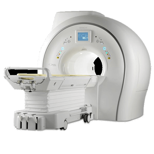
West Ashley
Weight limit 550 lbs, bore width 74cm
GE 3T Discovery Wide Bore
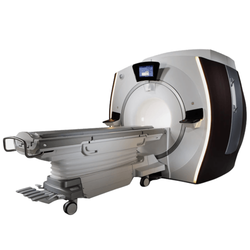
Tricom
Weight limit 500 lbs, bore width 70cm
GE 1.5T Signa HD
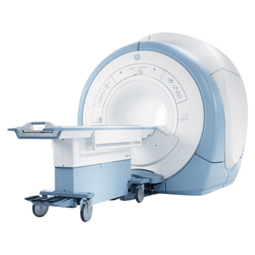
Tricom
Weight limit 350 lbs, bore width 54cm
Physician Links
Patient Links
Get To Know Us
Interpretation services
Get To Know Us
Patient Links
Physician Links
Interpretation services
© Tricounty Radiology, All Rights Reserved
Translation services on this website are provided via Google™ Translate, a free automated translation service that can translate text into different languages. This tool is for your convenience only, and should not be considered exact and may in some cases include incorrect language. No warranty of any kind is made as to the accuracy, correctness, or reliability of any information translated by Google™ Translate. Please know that when a translation is requested, you will be leaving the the Tricounty Radiology website and any person or entity who relies on these translation services does so at his or her own risk. If you have any questions about Google™ Translate, please click the following link: Google™ Translate FAQs.